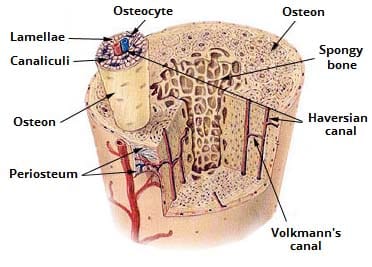
Compact Bone Diagram Canaliculi - Figure 6 1 The Bones And Cartilages Of / Most of the lamellae of compact bone are organized into sets of concentric rings with each set surrounding a central, or haversian, canal.
byAdmin-
0
Compact Bone Diagram Canaliculi - Figure 6 1 The Bones And Cartilages Of / Most of the lamellae of compact bone are organized into sets of concentric rings with each set surrounding a central, or haversian, canal.. The compact bone is the main structure in the body for support, protection, and movement. Compact bone compact bone is the denser stronger of the two types of bone tissue figure 6. They allow blood vessels and nerves to travel through them to supply the osteocytes. There are two types of bone tissue: The osteocytes sit in their lacunae in concentric rings around a central haversian canal (which runs longitudinally).
Most of the lamellae of compact bone are organized into sets of concentric rings with each set surrounding a central, or haversian, canal. (b) in this micrograph of the osteon, you can clearly see the concentric lamellae and central canals. Compact bone appears to be solid while spongy bone has the appearance of a sponge. Anatomy of a long bone proximal epiphysis diaphysis distal epiphysis compact bone spongy bone medullary cavity. Composed of compact bone and spongy bone.
Periosteum Png Images Pngegg from e7.pngegg.com It is thick and dense. The bone contains a multitude of small irregular spaces, approximately fusiform in shape, called lacunae, with very minute canals leading from them and anastomosing with similar little prolongations from the other lacunae. Bone canaliculi are microscopic canals between the lacunae of ossified bone. Anatomy of a long bone proximal epiphysis diaphysis distal epiphysis compact bone spongy bone medullary cavity. Describe the structure of compact bone. The canaliculi are small channels that link together the lacunae as well as having a function of routing nutrients to osteocytes and expelling waste products. Compact bone, as opposed to spongy bone, is made of cylindrical units, called osteons, that are tightly formed together. It is also called osseous tissue or cortical bone and it provides structure and support for an organism as part of its skeleton, in addition to being a location for the storage of minerals like calcium.about 80% of the weight of the human skeleton comes from.
Due to the strong nature of compact bone, compared to spongy bone, it is the preferred tissue for strength.
Compact bone is the denser stronger of the two types of bone tissue. Each central canal, with the Compact bone, also called cortical bone, dense bone in which the bony matrix is solidly filled with organic ground substance and inorganic salts, leaving only tiny spaces (lacunae) that contain the osteocytes, or bone cells.compact bone makes up 80 percent of the human skeleton; Shown is a longitudinal section from the human ulna, showing haversian canal, lacunae, and canaliculi. Describe the structure of compact bone. Good, here in this part, i am going to describe the structure of compact bone. Haversian canals (sometimes canals of havers) are a series of microscopic tubes in the outermost region of bone called cortical bone. The canaliculi are small channels that link together the lacunae as well as having a function of routing nutrients to osteocytes and expelling waste products. However, compact bones also serve a function in storing and releasing calcium to the. Diagram of a typical long bone showing both cortical (compact) and cancellous (spongy) bone. Most long bones that need to lengthen rapidly form this way due to the ability of hyaline cartilage to form quickly without requiring a direct blood supply. The compact bones form the hard exterior of the bones, whereas the spongy bones have several pores that are filled with nerves and blood vessels. About press copyright contact us creators advertise developers terms privacy policy & safety how youtube works test new features press copyright contact us creators.
The remainder is cancellous bone, which has a spongelike appearance with numerous large spaces and is found in the. (b) in this micrograph of the osteon, you can see the concentric lamellae around the central canals. Osteon model lacunae canaliculi osteocyte. This type of bone is located between layers of compact bone and is thin and porous. Compact bone also called cortical bone dense bone in which the bony matrix is solidly filled with organic ground substance and inorganic salts leaving only tiny spaces lacunae that contain the osteocytes or bone cells.
Bone Tissue Amboss from media-us.amboss.com Diagram of a typical long bone showing both cortical (compact) and cancellous (spongy) bone. Good, here in this part, i am going to describe the structure of compact bone. Learn vocabulary, terms, and more with flashcards, games, and other study tools. The compact bones form the hard exterior of the bones, whereas the spongy bones have several pores that are filled with nerves and blood vessels. Compact bone, or cortical bone, mainly serves a mechanical function. Some, mostly older, compact bone is remodelled to form these haversian systems (or osteons). Collaborative flowcharts, wireframes, mind maps and sticky notes. Describe the structure of compact bone.
Compact bone appears to be solid while spongy bone has the appearance of a sponge.
The remainder is cancellous bone, which has a spongelike appearance with numerous large spaces and is found in the. They allow blood vessels and nerves to travel through them to supply the osteocytes. Spongy bone is used for more active functions of the bones, including blood cell production and ion exchange. The compact bones form the hard exterior of the bones, whereas the spongy bones have several pores that are filled with nerves and blood vessels. The canaliculi are small channels that link together the lacunae as well as having a function of routing nutrients to osteocytes and expelling waste products. There are two types of bone tissue: Haversian canals (sometimes canals of havers) are a series of microscopic tubes in the outermost region of bone called cortical bone. Haversian canal, lacuna, circumferential lamella, interstitial lamellae, osteocyte, canaliculi, matrix, osteon 2. Lacunae are minute spaces that contain bone cells, otherwise known as the osteocytes. (b) in this micrograph of the osteon, you can clearly see the concentric lamellae and central canals. (b) in this micrograph of the osteon, you can clearly see the concentric lamellae and central canals. Do you want to learn the details of the histology of compact bone with labelled diagram and authentic slide images? Anatomy of a long bone proximal epiphysis diaphysis distal epiphysis compact bone spongy bone medullary cavity.
The canaliculi are small channels that link together the lacunae as well as having a function of routing nutrients to osteocytes and expelling waste products. Use the venn diagram to compare and contrast compact bone and spongy bone. The compact bones form the hard exterior of the bones, whereas the spongy bones have several pores that are filled with nerves and blood vessels. This type of bone is located between layers of compact bone and is thin and porous. It is thick and dense.
Ultrastructure Of Bone Components Structure Teachmeanatomy from teachmeanatomy.info Compact bone, or cortical bone, mainly serves a mechanical function. There are two types of bone tissue: Compact bone appears to be solid while spongy bone has the appearance of a sponge. The canaliculi are small channels that link together the lacunae as well as having a function of routing nutrients to osteocytes and expelling waste products. Anatomy of a long bone proximal epiphysis diaphysis distal epiphysis compact bone spongy bone medullary cavity. Compact bone, as opposed to spongy bone, is made of cylindrical units, called osteons, that are tightly formed together. About press copyright contact us creators advertise developers terms privacy policy & safety how youtube works test new features press copyright contact us creators. This is the area of bone to which ligaments and tendons attach.
The canaliculi are small channels that link together the lacunae as well as having a function of routing nutrients to osteocytes and expelling waste products.
Compact bone histology slide structure with diagram. The canaliculi are small channels that link together the lacunae as well as having a function of routing nutrients to osteocytes and expelling waste products. Start studying compact bone under microscope. It is also called osseous tissue or cortical bone and it provides structure and support for an organism as part of its skeleton, in addition to being a location for the storage of minerals like calcium.about 80% of the weight of the human skeleton comes from. Most of the lamellae of compact bone are organized into sets of concentric rings with each set surrounding a central, or haversian, canal. Haversian canal, lacuna, circumferential lamella, interstitial lamellae, osteocyte, canaliculi, matrix, osteon 2. Good, here in this part, i am going to describe the structure of compact bone. (b) in this micrograph of the osteon, you can clearly see the concentric lamellae and central canals. Compact bone is the denser stronger of the two types of bone tissue. Bone formation from the cartilage mold. Concentric lamellae interstitial lamellae central canal lacuna osteocyte canaliculus. The compact bone is the main structure in the body for support, protection, and movement. They allow blood vessels and nerves to travel through them to supply the osteocytes.
(b) in this micrograph of the osteon, you can see the concentric lamellae around the central canals compact bone diagram. Osteon model lacunae canaliculi osteocyte.